| Architecture of 3DNA Dendrimer | |
| What is a dendrimer? The name “Dendrimer”, derived from the Greek word for “tree”, suggests the unusual structure of this highly branched molecule. As a class, dendrimers are complex, branched molecules built from interconnected natural or synthetic monomeric subunits. A 3DNA dendrimer is constructed from DNA monomers – as the name “3DNA” indicates. 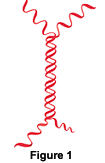 The 3DNA monomer - the basic unit of dendrimer construction Each 3DNA monomer is composed of two DNA strands that share a region of sequence complementarity located in the central portion of each strand. When the two strands anneal to form the monomer the resulting structure has a central double-stranded “waist” bordered by four single-stranded “arms” [Figure 1]. This “waist” plus “arms” structure comprises the basic 3DNA monomer. Linking monomers - the architecture of a 3DNA dendrimer The single-stranded “arms” at the ends of each of the five monomer types are designed to interact with one another in precise and specific ways. Base-pairing between the “arms” of complementary monomers allows directed assembly of the dendrimer as a step-wise series of monomer layers [Figures 2A to 2C].    The layered structure of the 3DNA dendrimer Construction of a 3DNA dendrimer begins with a single initiator monomer. To this, a first layer of monomers is attached by annealing the “arms” of the first layer monomers to the “arms” of the initiator monomer. The result is a one-layer 3DNA dendrimer with 12 single-strand “arms” available on its surface [Figure 2A]. This structure is then chemically crosslinked to prevent dissociation. Next, a second layer of monomers is attached to the first layer using the same interaction between the single-stranded “arms” of each component. In this two-layer 3DNA dendrimer the number of free single-stranded “arms” increases to 36 [Figure 2B]. A third layer of monomers is added in an analogous fashion, creating a total of 108 free single-stranded arms. Finally, in a perfect four-layer dendrimer, addition of the last set of monomers leaves 324 singlestranded “arms” on the surface of the molecule [Figure 2C]. As the dendrimer manufacturing process is not perfect, most dendrimers have fewer than 324 arms. An average dendrimer has about 250 arms, has a diameter of 150-200 nanometers, and consists of about 36,000 bases of DNA. Using the “arms” for two functions – labeling and application specificity The “arms” on the surface of the 3DNA dendrimer are used to attach the dendrimer’s two key functionalities. One function of the arms is to enable attachment of label. The other is to make the dendrimer specific to a particular application or experiment.  3DNA functionalities are attached with oligonucleotides The molecules that determine the target and labeling specificity of the dendrimer are attached either as oligonucleotides or as oligonucleotide conjugates. Using simple DNA labeling, hybridization and ligation reactions, the 3DNA dendrimer scaffold may be converted into a highly labeled, applicationspecific probe [Figures 3A to 3C].   Flexible labeling and specificity The architecture of the 3DNA dendrimer imposes no restrictions on the type of specificity or label used, so the dendrimer may be configured to meet a wide variety of detection needs. The label may be fluorescent, enzymatic, or radioactive. The application specificity may take the form of a generic “capture sequence”, as in our Array 900 kits, of a targetspecific sequence, or of an antibody, as in our UltraAmp reagents. Passive signal enhancement Signal intensity is determined by the amount of label that can be localized at the reaction site. The 3DNA dendrimers in our kits are labeled with an average of at least 200 labels (more in some kits). The dendrimer carries this number of labeles with it every time it binds to a complementary molecule. The result is up to a 200-fold passive enhancement of signal intensity. |
|
| Articles: Nilsen,T.W., Grazel, J., Prensky,W., Dendritic Nucleic Acid Structures, J. Theoretical Biology, 187:273-284 (1997). Capaldi, S., Getts, R.C., and Jayasena, S.D., A Signal Amplification Through Nucleotide Extension and Excision on a Dendritic DNA Platform, Nucl. Acids Res., 28(7):21e (2000). Wang, J., Jiang, M., Nilsen, T. W., and Getts, R., Dendritic Nucleic Acid Probes for DNA Biosensors. J. Am. Chem. Soc., 120:8281-8282 (1998). Wang, J., Rivas, G., Fernandes, J., Jiang, M., Lopez Paz, J.L., Waymire, R., Nilsen, T. W., and Getts, R., Adsorption and Detection of DNA Dendrimers at Carbon Electrodes., Electroanalysis, 10(8):553-556 (1998). |
| Array Detection | RNA Amplification | About 3DNA | Signal Amplifiers Literature | Tech Support | Distributors | Newsletters | Contact Us Copyright © 2006 Genisphere. All rights reserved. |